Course Navigation
Course Home Expand All

Rock Fracture Under Confined Compression

Shear vs Tensile Failure

Rock has two primary modes of failure that vary depending on the stress conditions and the intrinsic rock strength. Hydraulic fracture is a tensile mode of failure, where the relative displacement between the fracture faces or surfaces is opening. Shear failure is the second predominant mode, where the relative displacement between the two fracture faces is sliding.

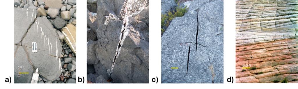
Tensile (also called opening mode) fractures in the subsurface typically grow when the fluid pressure in the rock pores exceeds the minimum compressive stress in the rock, causing the walls of pre-existing microcracks to open relative to each other. If the fracture strength of the material is exceeded, the crack tips will propagate. Preserved natural hydraulic fractures in the rock record show evidence of crack opening in features called veins, that have mineral fill that props the fracture opening, and in joints, which have barren fracture walls that may still be opened or may have closed upon stress relaxation (Figure 3.3.1).

Figure 3.3.1: Natural tensile fractures in rock. a) Calcite-filled veins in limestone. Bristol Channel, England. b) Partially mineralized quartz vein in quartzite, showing preserved porosity. Ozark Mountains, Arkansas. c) Open joints in limestone. Pedernales Falls State Park, Texas. d) Nominally closed joints. Checkerboard Mesa, Arches National Park, Utah.
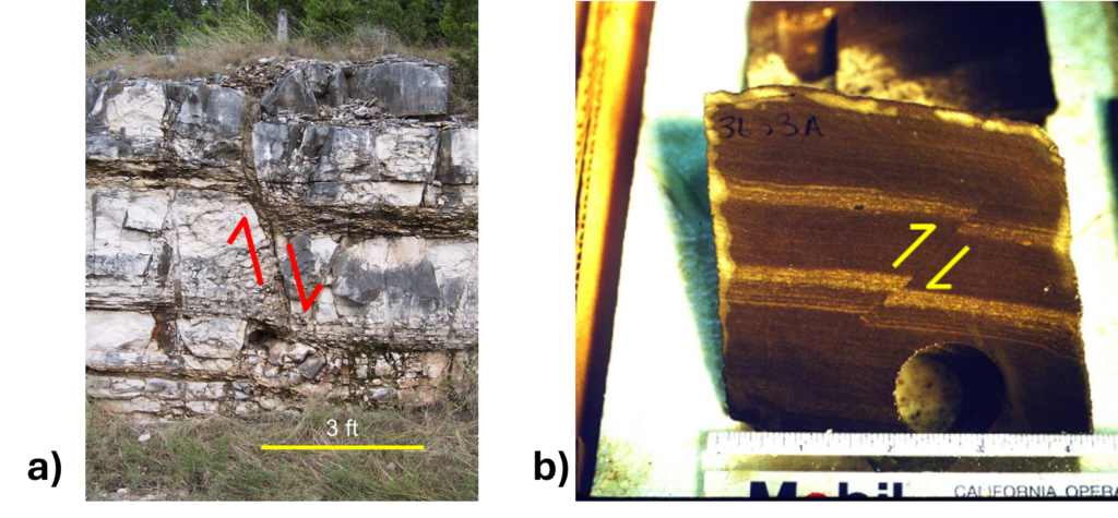
Natural shear fractures in rock are called faults. The relative shear displacement created by sliding can be detected by the offset of markers in the rock such as bedding planes or fossils.

Figure 3.3.2: Faults. a) A normal fault (due to horizontal extension) in a limestone outcrop near New Braunfels, Texas. Fault offset is approximately 1 ft. b) A reverse fault (due to horizontal shortening) in mudstone from the South Ellwood Field, offshore California. Sample is a slab-cut vertical core that is 4 inches in diameter. Fault offset is approximately 1 cm.


Rock fracture photos in Figures 3.3.1 and 3.3.2 were taken by Jon E. Olson, Chair Emeritus, The University of Texas at Austin.